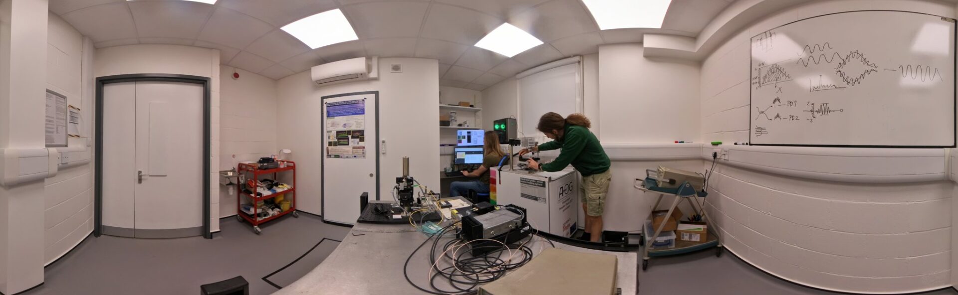
We develop imaging and sensing systems primarily for medical and biomedical applications, but also for industry, scientific research and conservation science.
The Applied Optics Group has expertise across a wide range of optical imaging modalities, including optical coherence tomography (OCT), microscopy and photoacoustic imaging.
Our work ranges from developing new components, particularly optical sources, through novel software and processing approaches (including artificial intelligence), to full system development and translational studies.
Examples of current and recent externally-funded projects include:
- FoVEnOCT – A three-year EPSRC-funded project to develop compact forward-viewing OCT probes for endoscopic use using a technique patented by Kent.
- OCT-Raman – Quantitative OCT-Raman spectral imaging for intra-operative detection of positive margins in breast conserving surgery, an MRC-funded collaboration with University of Nottingham [Press Release].
- VIPER – Sensorised Micro-Surgical Robot for Safe Regenerative Retinal Therapy Delivery, an NIHR funded collaboration with King’s College London to augment retinal surgical robots with distance sensing and imaging. [Press Release]
- An Optical Detector For Latent Fungal Infection In Produce – a BBSRC-funded collaboration with NIAB East Malling to develop OCT imaging for fruit.
- 5DHiResE – a BBSRC-funded project to achieve 5-dimensional high-resolution non-invasive assessment of mammalian embryos.
- REBOT – Robotic endobronchial optical tomography, an EPSRC-funded collaboration with Imperial College London to develop robot-guided imaging probes for the lung.
- NETLAS – An EU-funded doctoral training school on Next generation of tunable lasers for optical coherence tomography, co-ordinated by the AOG.
More details of our current and previous funded projects are available on the Research Projects page. A list of publications can be found on the Publications page.
Optical coherence tomography
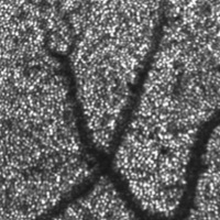
Photo-receptors visible on the retina
Optical coherence tomography (OCT) is a non-invasive, high resolution optical imaging technique, widely used for imaging the retina, but with many other emerging medical and industrial applications. The AOG was at the forefront of developments in en face OCT and combining OCT with scanning laser ophthalmoscopy, and more recently invented the master-slave OCT technique for high-speed processing of raw Fourier domain data.
Our research includes adaptive optics for enhanced resolution as well as techniques for simultaneous multi-depth imaging, dynamic focus, phase sensitive OCT, polarisation-sensitive OCT, full-field OCT, and speckle reduction. Developments also include new swept sources for Fourier domain imaging. In collaboration with NKT Photonics, via a European Industrial Doctorate (UBAPHODESA), we recently worked on large bandwidth optical sources and their applications for multi-modal optical imaging and we co-ordinated NETLAS, a European Marie Curie Training School developing next generation tunable lasers for optical coherence tomography.
Applications of optical coherence tomography
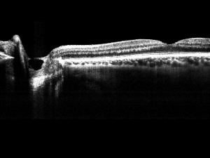
Optical coherence tomography cross-section through the human retina.
We have worked with a wide range of collaborators to develop ophthalmic and wider medical and industrial applications of OCT, including to areas such as embryology, lung disease, and art conservation. We have also contributed to the developed of non-medical applications such as art conservation and forensic sciences. Our systems have been deployed to several hospitals for clinical trials, including Northwick Park Hospital in London and as far afield as the New York Eye and Ear Infirmary.
Multi-modal optical imaging
We have combined OCT and other imaging techniques to achieve multi-modality biomedical imaging. The first combined scanning laser ophthalmoscope (confocal microscopy for the eye) and optical coherence tomography (SLO/OCT) was patented at Kent by Adrian Podoleanu and David Jackson in 1998 (US Patent 5975697). The technology was exclusively licensed to Ophthalmic Technology Inc (OTI), Canada. Work at Kent later added a fluorescence channel to the system, allowing simultaneous OCT, SLO and indocyanine green (ICG) fluorescence imaging of the retina. We have also used combined fluorescence and OCT imaging for studying specimens such as Drosophila melanogaster larval heart. Recently we have been working to combine photoacoustic imaging with OCT using high-energy supercontinuum sources from NKT Photonics.
Handheld and endoscopic imaging
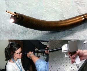
Endoscopic OCT systems
We have developed a research programme in miniaturised handheld and endoscopic OCT and fluorescence imaging systems for minimally invasive medical diagnostics. For example, in collaboration with Northwick Park Hospital and the Institute of Applied Physics in Russia, we developed a forward-viewing OCT probe combined with a fibre bundle endoscope for dual-mode ear, nose and throat (ENT) imaging and worked on robot-guided OCT and fluorescence imaging probes for the lung, as part of the REBOT project in collaboration with Imperial College London. We are also working on endoscopic and needle-based fluorescence microscopes as well as low-cost and handheld OCT probes.
Optical measurement and sensing
The AOG has a long history in optical sensing and measurement for example in structural health monitoring, distributed sensing, metrology, and medical sensing, as well as in high-speed microwave photonics and optical correlators. Recently, we demonstrated a coherence-gated wavefront sensor for depth-resolved measurements of wavefront aberrations.