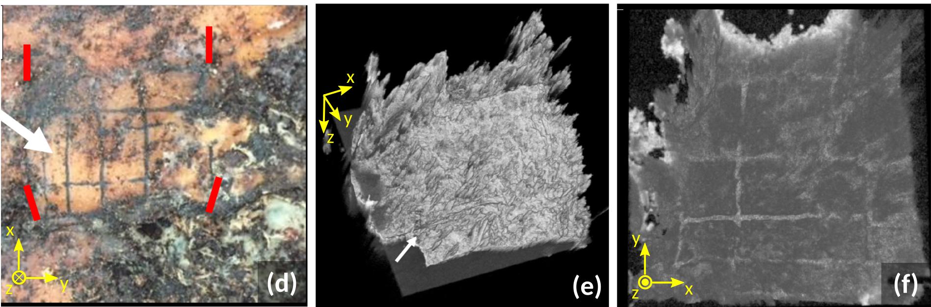
(image above: OCT volumes from decomposed porcine tissue, tattooed post-mortem, from doi:10.1117/12.2526757.)
Poster presented at ECBO, on recognising tattooed patterns in decomposing tissue. Click to enlarge.
Optical Coherence Tomography (OCT) has become one of the most successful and significant techniques in clinical, non-invasive measurements, particularly in the opthalmic field. OCT provides three-dimensional sample visualisation, using visible and infra-red light to penetrate sub-surface into samples. While being primarily used in the biomedical fields (ophthalmic, dermal, endoscopic), it can be equally applied to non-destructive testing, in art conservation, quality control, and forensic sciences.
The Applied Optics Group, with its long history of developing OCT systems and applications (spanning more than two decades), has also been engaging with a range of stakeholders in order to further develop the range of applications in forensic science, through a strong collaboration with Robert Green, OBE, Senior Lecturer in Forensic Science at the School of Physical Sciences. In particular, two research lines have been explored recently:

A volume obtained via OCT imaging of a security feature in a Portuguese National ID card.
OCT and forensic anthropology
Through a collaboration with Dr Chris Deter, from the School of Anthropology and Conservation at the University of Kent, we have researched the feasibility of OCT imaging to recognise tattooed patterns in decomposing tissue (porcine skin). Tattoos (and other skin markings) are useful secondary means of identification, employed in Disaster Victim Identification (DVI) when no primary means of identification (eg. DNA, dental records) are available for comparison. This work yielded a conference (poster) presentation at the European Conferences in Biomedical Optics in Munich, Germany.
OCT and forensic document examination
More recently, we have been investigating the feasibility of OCT imaging in forensic document examination, namely in assessing the authenticity of identification documents by analysing security features which may be located sub-surface in the document (eg. holograms, multi-layered structures). For this work, we have been advised by industry partners, namely those at Foster and Freeman, Inc.
This collaboration has culminated on the publication of a peer-reviewed article in Science and Justice (the journal of The Chartered Society of Forensic Sciences).
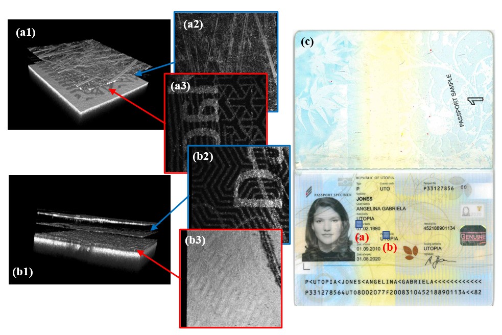
Virtual “dissection” of a modern polycarbonate specimen passport, showing different security features. Volumes (a) and (b), each approximately 2×2 mm2, were taken from a specimen polycarbonate passport (sub-figure (c)). The 3-D volume renders in sub-figures (a1) and (b1) showcase the different layers in the passport, with the corresponding en-face slices (a2)-(a3) and (b2)-(b3) shown alongside them.