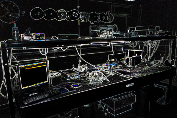
With previous support from an EPSRC First Grant and a Vice Chancellor’s PhD Studentship we have been developing miniaturised endoscopic and needle microscopes for minimally-invasive medical diagnostics and interventional guidance.
Optical biopsy (or endomicroscopy) is a real-time, minimally-invasive alternative to conventional histology, offering the prospect of faster and more accurate medical diagnostics and image-guided intervention. The idea is to acquire microscopy images of tissue in situ using a probe-based microscope. One way of doing this is to use a thin, flexible fibre imaging bundle to relay an image from the tissue to an external microscope. This avoids the need for any electronics or scanning systems to be built into the probe, allowing it to be thinner and less invasive, and allowing it to be passed down the narrow working channels of endoscopes. Nevertheless, fibre bundles do have their limitations, particularly in terms of the resolution and field-of-view of the microscope images. At Kent we are working on developing lower-cost and more capable endomicroscopes, with the aim of facilitating new applications, both in developed and low-resource settings.
Needle Microscopes (EPSRC Project)
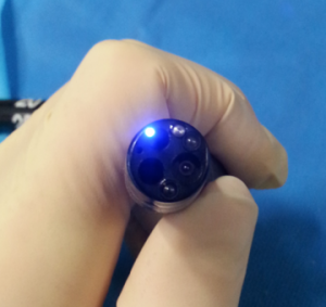
We are working on developing thinner endomicroscopes which can be used as needle probes – penetrating beneath the surface of the tissue. The current generation of needle probes, using fibre image bundles or gradient index lenses as image conduits, suffers from poor resolution and a low pixel count, limiting the range of potential applications. We are working on new techniques for imaging through ultra-narrow conduits, with the aim of improving the resolution and field-of-view of the images, while simultaneously reducing the outer diameter of the probe. This involves numerical simulations, designing, building and testing optical/electrical hardware and software, and developing clinical applications together with collaborators.
For more information on projects involving endoscopic and needle microscopes, please see Dr Mike Hughes lab or contact by by email.
Selected paper authored or co-authored by Michael Hughes on endomicroscopy:
A Thrapp and M. Hughes, “Reduced motion artifacts and speed improvements in enhanced line-scanning fiber bundle endomicroscopy,” Journal of Biomedical Optics, 26 (5) (2021) https://kar.kent.ac.uk/88084/
M. Hughes, “Inline holographic microscopy through fiber imaging bundles.” Applied Optics, 60 (4). A1-A7. https://kar.kent.ac.uk/83241/
A Thrapp and M. Hughes, “Automatic motion compensation for structured illumination endomicroscopy using a flexible fiber bundle,” Journal of Biomedical Optics, 25 (2). (2020) [Open Access]
K. Vyas, M. Hughes, B. Rosa and G-Z. Yang, “Fiber bundle shifting endomicroscopy for high-resolution imaging,” Biomedical Optics Express 9(10), 4649 (2018). [Open Access].
P. Giataganas, M. Hughes, C. Payne, P. Wisanuvej, B. Temelkuran, and G-Z. Yang. “Intraoperative robotic-assisted large-area high-speed microscopic imaging and intervention.” IEEE Transactions on Biomedical Engineering (2018).
A.J. Thompson*, M. Hughes*, S. Anastasova, L.S. Conklin, T. Thomas, C. Leggett, W.A. Faubion, T. J. Miller, P. Delaney, F. Lacombe, S. Loiseau, A. Meining, R. Richards-Kortum, G. J. Tearney, P. Kelly & G-Z. Yang, “The potential role of optical biopsy in the study and diagnosis of environmental enteric dysfunction,” Nature Reviews Gastroenterology and Hepatology, Advance Publication (2017). [* Joint First Author].
L. Zhang, M. Ye, P. Giataganas, M. Hughes, A. Bradu, A. Podoleanu, G-Z. Yang, “From Macro to Micro: Autonomous Multiscale Image Fusion for Robotic Surgery,” Robotics and Automation Magazine 24(2), 63-72 (2017). [Open Access].
S. Zuo, M. Hughes, G-Z. Yang, “Flexible Robotic Scanning Device for Intraoperative Endomicroscopy in MIS,” IEEE Trans. Mechatronics 2017 [Early Access].
K. Vyas, M. Hughes, D. Leff, G-Z. Yang, “Methylene-blue aided rapid confocal laser endomicroscopy of breast cancer,” J. Biomed. Opt. 22(2), 020501 (2017) [PDF]
M.Hughes, G-Z.Yang, “Line-scanning fiber bundle endomicroscopy with a virtual detector slit,” Biomedical Optics Express 7(6), 2257-68 (2016). [PDF]
S. Zuo, M. Hughes, G-Z.Yang, “Novel balloon surface scanning device for intraoperative breast endomicroscopy,” Annals of Biomedical Engineering 62(12) 2941-52 (2015).
T. Chang, D. Leff, S. Shousha, D. Hadjiminas R. Ramakrishnan, M. Hughes, G-Z.Yang, A. Darzi, “Imaging breast cancer morphology using probe-based confocal laser endomicroscopy: Towards a real-time intraoperative imaging tool for cavity scanning,” Breast Cancer Research and Treatment 153(2), 299-310 (2015).
S. Zuo, M. Hughes, C. Seneci, T.P. Chang, G-Z.Yang, “Towards intraoperative breast endomicroscopy with a novel surface scanning device,” IEEE Transactions on Biomedical Engineering 62(12) 2941-52 (2015).[PDF]
P. Giataganas, M. Hughes, and G-Z.Yang, “Force adaptive robotically assisted endomicroscopy for intraoperative tumour identification,” International Journal of Computer Assisted Radiology and Surgery (2015) (Runner Up Best Paper at IPCAI 2015.) [Pre-print PDF]
M. Hughes and G-Z. Yang, “High speed, line-scanning, fiber bundle fluorescence confocal endomicroscopy for improved mosaicking,” Biomedical Optics Express 6, 1241-1252 (2015). [Open Access]
M. Hughes, P. Giataganas, and G-Z. Yang, “Color reflectance fiber bundle endomicroscopy without back-reflections,” Journal of Biomedical Optics 19(3), 030501 (2014). [PDF]
M. Hughes, T. P. Chang and G-Z. Yang, “Fiber bundle endocytoscopy,” Biomedical Optics Express 4(11) 2781-94 (2013) [PDF]