Above: preliminary results from a past publication (Caujolle et al., Biomed Opt Express 8(11) (2017), doi:10.1364/BOE.8.005139) where bovine embryos were imaged in-situ.

The team currently behind the 5DHiResE project is reseaching the use of non-invasive, non-destructive imaging techniques, such as Optical Coherence Tomography (OCT) to establish the vitality of in-vitro produced (IVP) embryos (both bovine and porcine). This project is the result of a cross-disciplinary collaboration between the AOG and the Prof Griffin’s lab in the School of Biosciences. As partner industrial organisations, this project is reliant on the support of ActIVF-ET, Care Fertility Group, and Topigs Norsvin.
More preliminary results
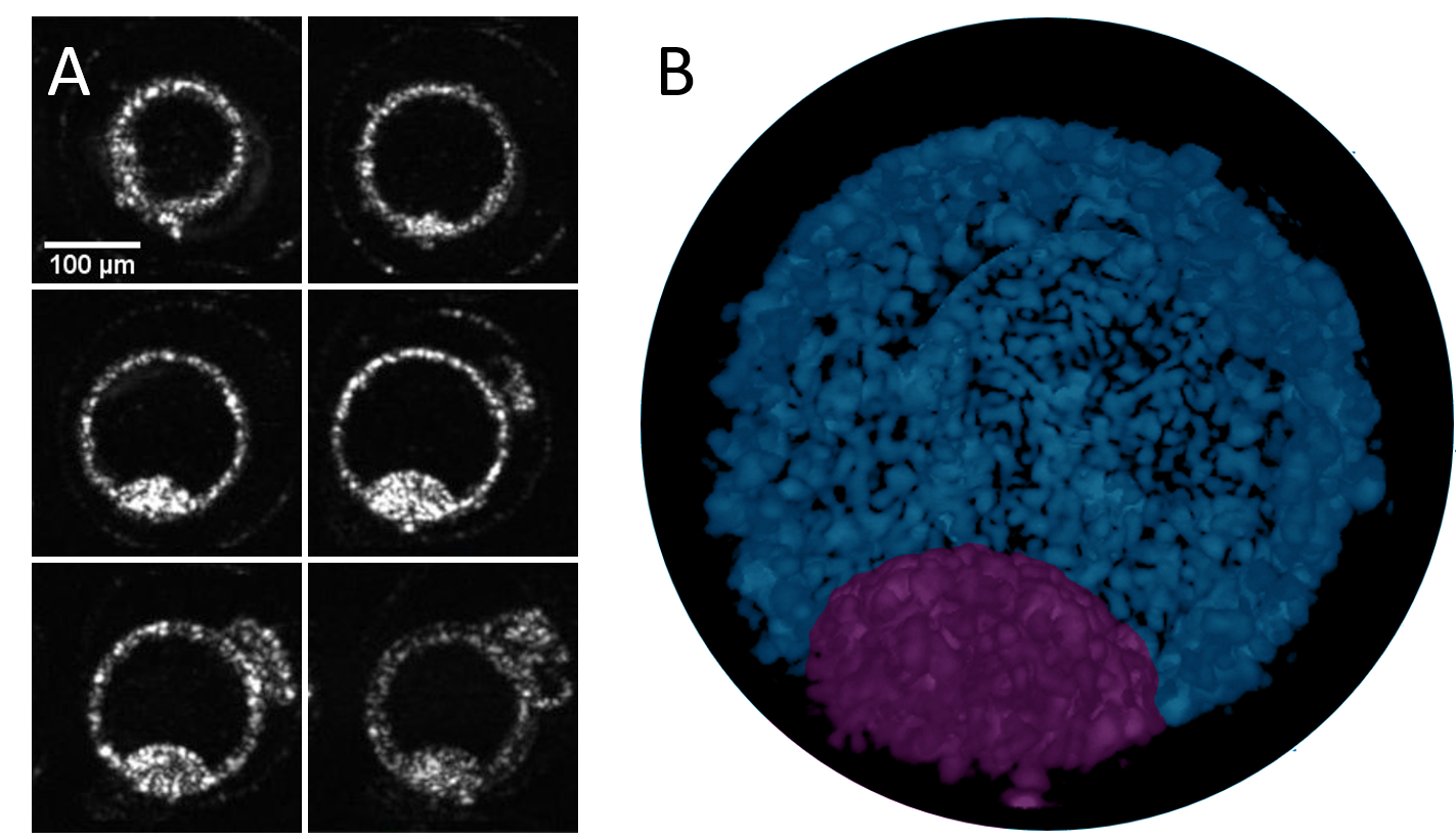
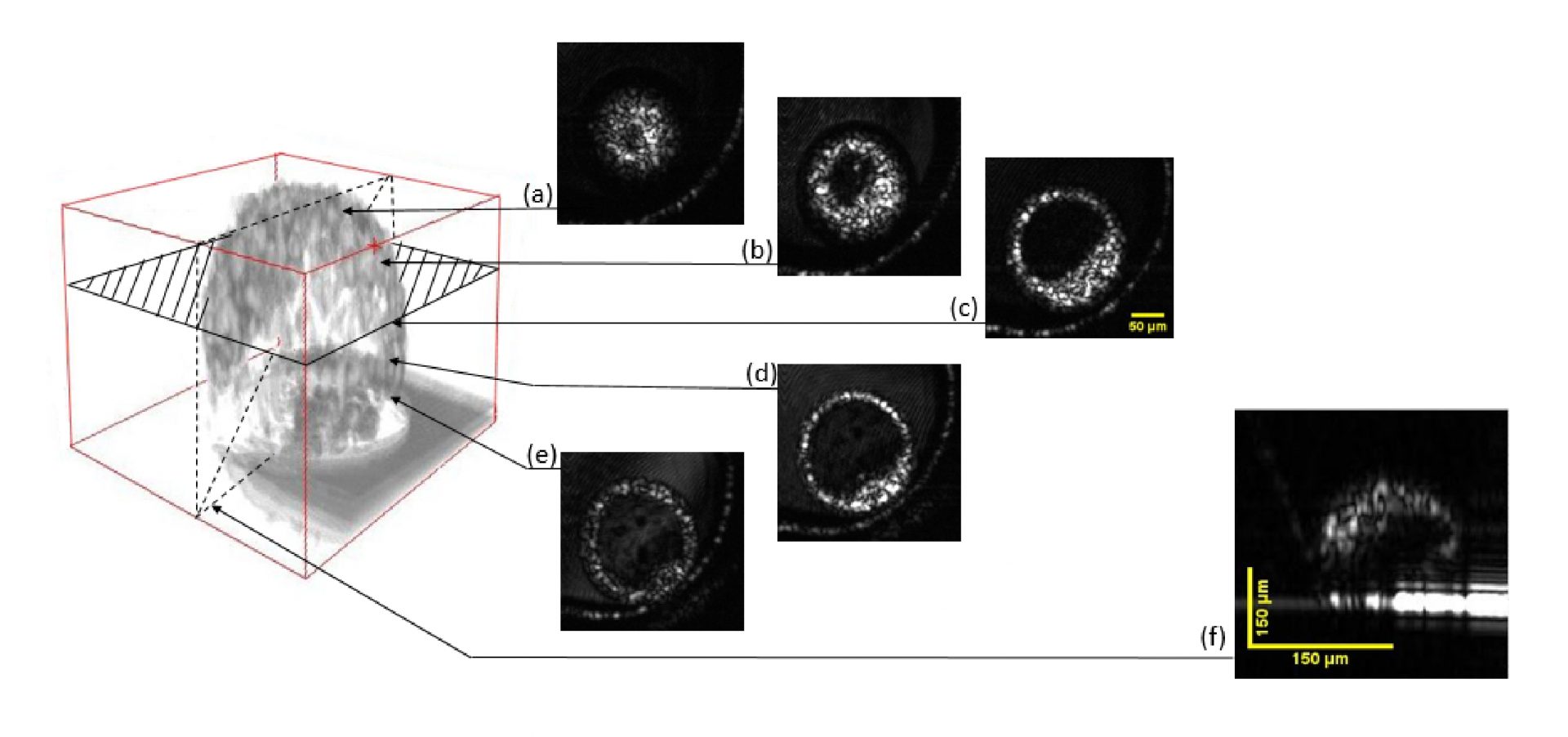
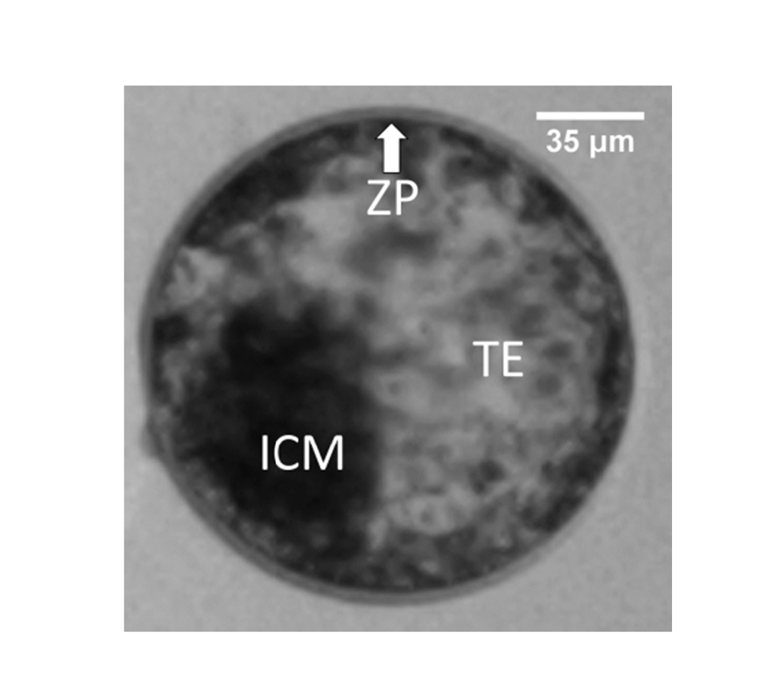
OCT reconstruction of a bovine blastocyst at day 7 of development. A) A selection of en-face images captured at different depths in the embryo. B) 3D model of the embryo reconstructed on ImageJ from approximately 300 en-face images. For display purposes, a window was created to allow the visualisation of the inside of the embryo and artificial colours were applied to the future placenta (trophectoderm, blue) and the future foetus (inner cell mass, purple). A rotating model of the same embryo can be seen in the animated GIF above.