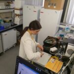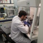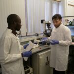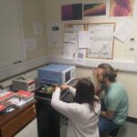
Optical coherence tomography (OCT) is an optical method for the 3D study of a sample. Its application to embryology could improve our understanding of early human/animal development, advancing embryo selection tools for IVF (in-vitro fertilisation). Recently, the Applied Optics Group developed a novel, infrared light-powered OCT imaging system (as part of the 5DHiResE project). This project aims to employ porcine semen as a model to assess the safety of this equipment. Sperm samples will be exposed to the light source and their motion, DNA and membrane integrity compared against appropriate controls, requiring an interdisciplinary team: one controlling/varying the illumination conditions and another evaluating the samples post-exposure.
OCT is a novel ‘optical biopsy’ method for obtaining cross-sectional 3D live images and its application to embryology may assist in improving embryo selection. Our group has provided a proof-of-concept for this method [1] and has recently reported the development of the first OCT time-lapse system, employing an infrared laser source [2]. However, an adequate investigation into the potential toxic effect OCT imaging systems might have in embryology is still needed – most guidance concerning exposure limits to light beams concern humans, and in particular, their retinal tissue and skin. Here, by using an established model reproductive cell line (porcine spermatozoa) [3], we will investigate the effects of the laser light and the OCT scanning protocol employed during imaging. This will be achieved by exposing the spermatozoa samples to several controlled light dose experiments, ranging from varying the optical power illuminating the sample, to changing the scanning protocol (allowing longer/shorter exposures for more defined/coarser images). The impact that novel OCT imaging modes, such as OCT Angiography (which allows quantification of the motion of the sperm samples, therefore yielding an estimate on their vitality), may have on the light dose during imaging will also be evaluated, as these modes require several images collected at the same location and therefore increase the light dose on the sample. For each sample stemming from the controlled light dose experiments, their health status will be evaluated, incorporating metrics such as sperm motion pattern and membrane/DNA integrity.
References
- Caujolle et al., Biomed Opt. Express 8(11) (2017), http://dx.doi.org/10.1364/BOE.8.005139;
- Marques et al., ECBO, Munich (2021), http://dx.doi.org/10.1117/12.2616149;
- Vollmer et al. Sci Rep 9(1) (2019) http://dx.doi.org/10.1038/s41598-019-50929-z.