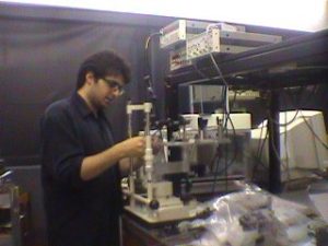
The combined OCT/SLO (optical coherence tomography and scanning laser ophthalmoscope) instrument for imaging the eye was invented at Kent in 1998 by Adrian Podoleanu and David Jackson.

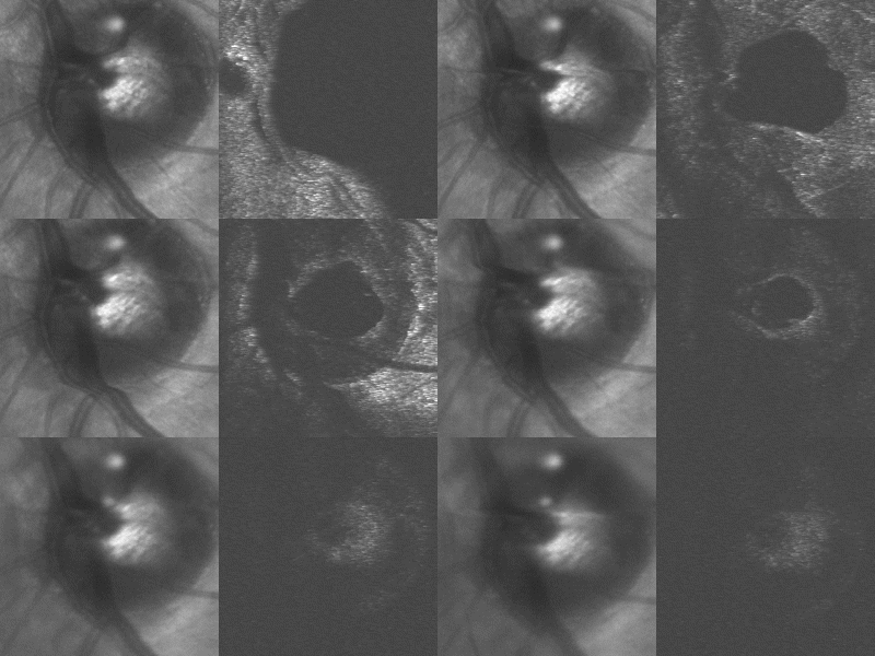
The output from a joint OCT/SLO system, with the SLO image on the left and the OCT on the right. The top of the optic nerve, then the RPE and at the end, the lamina cribrosa are seen in the OCT image. In the SLO image, these are visible all the time. The animation is made up from forty 3 mm x 3 mm images collected from the optic nerve in 20 seconds, with a total exploration depth of 1.5 mm.
- Optical mapping apparatus with adjustable depth resolution
The United States Patent 5975697 · Filed: 11/25/1998 · Published: 11/02/1999. - Combined Optical Coherence Tomograph and Scanning Laser Ophthalmoscope, A. Gh. Podoleanu and D. A. Jackson, Electronics Letters, Vol. 34, No. 11, (1998), pp. 1088-1090.
- Noise Analysis of a Combined Optical Coherence Tomograph and a Confocal Scanning Ophthalmoscope, A. Gh. Podoleanu, D. A. Jackson, Appl. Optics, Vol. 38, (1999), No. 10, pp. 2116 – 2127.
Our first OCT/SLO was an en-face system, with the OCT channel operating based on en-facetime domain OCT.

The assembly of a unique compact ophthalmological imaging system capable of en-face OCT/SLO as well as longitudinal SLO. This was a collaboration with Ophthalmic Technology Inc., Canada.
After 2002, spectral domain OCT technology took off, and the SLO channel was then paired sequentially with a spectral domain OCT channel. A system based on this principle was commercialised by Optos Plc., based on the patent:
The acronym OCT/SLO lives on, referring to spectral domain OCT which is now so fast that an en face fundus image can be created in real time from the OCT volume. Several groups have reported generation of these fundus images to guide conventional B-scan spectral domain OCT imaging, and these systems are now also described as OCT/SLO.
