During research on sensing between 1986-1995, the Applied Optics Group contributed towards several avenues which paved the way for the later development of what is known today as OCT. We were involved in all three types of OCT;
- Time domain OCT.
- Spectral or Fourier domain OCT, where the interferometer output is sent to an optical spectrometer.
- Swept source OCT, where a laser source is used which is swept within an equivalent band to that of the broadband source used in the time domain OCT or Fourier domain OCT.
A selection of papers published during that period:
- A. Gh. Podoleanu, S. Taplin, D. J. Webb, and D. A. Jackson, “Theoretical Study of Talbot-like Bands Observed Using a Laser Diode Below Threshold,” J. Pure and Applied Optics 7, 517-536 (1998).
- A. Gh. Podoleanu, S. Taplin, D. J. Webb and D. A. Jackson , “Talbot-like Bands for Laser Diode Below Threshold,” J. Pure and Applied Optics 6, 413 – 424, (1997).
- A. Gh. Podoleanu, S. Taplin, D. J. Webb and D. A. Jackson, “Channeled Spectrum Display using a CCD Array for Student Laboratory Demonstrations,” European J. Phys. 15, 266-271 (1994).
- A. Gh. Podoleanu, S. Taplin, D. J. Webb, and D. A. Jackson, “Channelled Spectrum Liquid Refractometer,” Rev. Sci. Instr. 64, 3028-3029 (1993).
- S. Taplin, A. Gh. Podoleanu, D. J. Webb, and D. A. Jackson, “Displacement Sensor Using Channeled Spectrum Dispersed on a Linear CCD Array,” Electron. Lett. 29, 896-897 (1993).
En-face flying spot OCT
For en face OCT, we showed that there is no need for an external phase modulator if the object to be imaged is scattering and the image size is sufficiently large. The modulation is interestingly, created by scanning the beam over the target:
- A. Gh. Podoleanu, G. M. Dobre, D. J. Webb, and D. A. Jackson, “Coherence Imaging by Use of a Newton Rings Sampling Function,” Opt. Lett. 21, 1789-1791 (1996).
Then we moved the centre of the Newton rings out from the image centre and sampled the target with a grid of line:
- A. Gh. Podoleanu, G. M. Dobre, and D. A. Jackson, “En-face Coherence Imaging Using Galvanometer Scanner Modulation,” Opt. Letters 23, 147-149 (1998).
This report demonstrates for the first time, B-scan OCT images from the retina constructed from T-scans (en-face 1D OCT scans):
- A. Gh. Podoleanu, M. Seeger, G. M. Dobre, D. J. Webb, D. A. Jackson, and F. Fitzke, “Transversal and Longitudinal Images from the Retina of the Living Eye Using Low Coherence Reflectometry,” J. Biomed. Opt 3, 12-20 (1998).
The OCT/SLO (collaboration with Ophthalmic Technologies Inc.)
The research on flying spot en-face OCT showed us how to produce OCT images with the same orientation as that in microscopy, or in scanning laser ophthalmoscopy (SLO). This allowed us to devise and assemble a dual imaging system. The system outputs pairs of OCT and confocal images. Several ophthalmoogy groups are now using the OCT/SLO for imaging the eye.
- A. Gh. Podoleanu, and D. A. Jackson, “Combined Optical Coherence Tomograph and Scanning Laser Ophthalmoscope,” Electron. Lett. 34, 1088-1090 (1998).
Collaboration with New York Eye and Ear Infirmary and the Department of Ophthalmology Academic Medical Center, University of Amsterdam :
- M. E. J. Van Velthoven, F. D.Verbraak, L. A. Yannuzzi, R. B. Rosen, A. Gh. Podoleanu, and Marc D. De Smet, “Imaging the Retina by En-Face Optical Coherence Tomography,” Retina 26, 129–136 (2006).
First OCT/ICG instrument for the eye, collaboration with New York Eye and Ear Infirmary:
- G. M. Dobre, A. Gh. Podoleanu, and R. B. Rosen, “Simultaneous optical coherence tomography–Indocyanine Green dye fluorescence imaging system for investigations of the eye’s fundus,” Opt. Lett., 30, 58-60, (2005).
Ultra high-resolution OCT/SLO system, collaboration with New York Eye and Ear Infirmary:
- R. G. Cucu, A.Gh. Podoleanu, J. A. Rogers, J. Pedro, and R. B. Rosen, “Combined confocal scanning ophthalmoscopy/en face T-scan based ultrahigh resolution OCT of the human retina in vivo,” Opt. Lett. 31, 1684-1687 (2006).
First OCT/SLO with AO correction, collaboration with National University of Ireland, Galway:
- R. G. Cucu, A. Gh. Podoleanu, J. A. Rogers, J. Pedro, and R. B. Rosen, “Combined confocal scanning ophthalmoscopy/en face T-scan based ultrahigh resolution OCT of the human retina in vivo,” Opt. Lett. 31, 1684-1687 (2006).
Sequential OCT/confocal. Sequential instead of simultaneous allows all signal to be used in each channel, OCT or confocal (SLO). (collaboration with New York Eye and Ear Infirmary):
- A. Gh. Podoleanu, G. M. Dobre, R. G. Cucu, and R. Rosen, “Sequential OCT and Confocal Imaging,” Opt. Lett. 29, 364-366 (2004).
Modulators and scanning methods for OCT
Using two RF modulators, we demonstrated simultaneous acquisition of two C-scans at two different depths:
- A. Gh. Podoleanu, G. M. Dobre, D. J. Webb, and D. A. Jackson, “Simultaneous En-face Imaging of Two Layers in Human Retina,” Opt. Lett. 22, 1039-1041 (1997).
Using a Mach Zehnder integrated modulator with independent RF excitation in each arm, we demonstrated simultaneous acquisition of two C-scans at two different depths, collaboration with University of Besancon:
- A. Gh. Podoleanu, J. A. Rogers, R. C. Cucu, D. A. Jackson, B Wacogne, H. Porte, and T. Gharbi, “Simultaneous Low Coherence Interferometry Imaging at Two Depths Using an Integrated Optic Modulator,” Opt. Comm. 191, 21-30 (2001).
Novel scanning delay line for fast A-scan but with less loss:
- C. C. Rosa, J. Rogers, and A. Gh. Podoleanu, “Fast Scanning Transmissive Delay Line Optical Coherence Tomography,” Opt. Lett. 30, 3263-3265 (2005)
Using two coupled interferometers we devised a novel method for measuring the eye length:
- A. Gh. Podoleanu, G. M. Dobre, D. J. Webb, and D. A. Jackson, “Fiberised set-up for Eye Length Measurement Full length,” Opt. Comm. 137, 397-405 (1997).
Corrections of distortions in OCT images
We make a distinction between scanning and refractive type distortions in OCT. This report also predicts a distorted elevation of the RPE layer in the fovea in the OCT images of the retina, due to differences in the indices of refraction of vitreous and retina (collaboration with University of Central Florida, School of Optics / CREOL and New York Eye and Ear Infirmary):
- A. Podoleanu, I. Charalambous, L. Plesea, A. Dogariu, and R. Rosen, “Correction of distortions in OCT imaging of the eye,” Phys. Med. Biol. 49, 1277-1294 (2004).
Signal to noise ratio analysis
Comparative noise analysis in the two channels of an OCT/SLO system:
- A. Gh. Podoleanu, and D. A. Jackson, “Noise Analysis of a Combined Optical Coherence Tomograph and a Confocal Scanning Ophthalmoscope,” Appl. Opt. 38, 2116 – 2127 (1999).
This report shows that fast OCT systems have to work in the excess photon noise regime limitation and not in the shot noise regime:
- A. Gh. Podoleanu, “Unbalanced versus balanced operation in an OCT system,” Appl. Opt. 39, 173-182 (2000).
Two novel noise bandwidth definitions are introduced to consider the excess photon noise in balanced OCT under wide bandwidth excitation:
- C. C. Rosa, and A. Podoleanu, “Limitation of the achievable signal to noise ratio in OCT due to mismatch of the balanced receiver,” Appl. Opt. 43, 4802-4815 (2004).
Applications of OCT
OCT in ophthalmology
Posterior pole. The majority of reports above referred to examples in imaging the retina. A novel method for topography using en-face OCT was presented in the following report (collaboration with Institute of Ophthalmology, London):
- J. A. Rogers, A. Gh. Podoleanu, G. M. Dobre, D. A. Jackson, and F. W. Fitzke, “Topography and volume measurements of the optic nerve using en-face optical coherence tomography,” Opt. Express 9, 476 – 545 (2001).
The utility of adjustable depth resolution required by en-face OCT method is presented in:
- A. Gh. Podoleanu, J. A. Rogers, and D. A. Jackson, “OCT En-face Images from the Retina with Adjustable Depth Resolution in Real Time,” IEEE J. Select. Top. Quant. Electron. 5, 1176-1184 (1999).
Anterior pole:
- A. Gh. Podoleanu, J. A. Rogers, G. M. Dobre, R. G. Cucu, and D. A. Jackson, “En-face OCT imaging of the anterior chamber,” Proc. SPIE 4619, 240-243 (2002).
OCT in imaging skin
- A. Gh. Podoleanu, J. A. Rogers, D. A. Jackson, and S. Dunne, “Three dimensional OCT images from retina and skin,” Opt. Express 7, 292-298 (2000).

An OCT/SLO pair of a carried tooth
OCT in dentistry
Collaboration with School of Dentistry, University of Liverpool:
- B. T. Amaechi, S. M. Higham, A. Gh. Podoleanu, J. A. Rogers, and D. A. Jackson, “Use of optical coherence tomography for assessment of dental carries: quantitative procedure,” J. Oral Rehab. 28, 1092-1093 (2001).
- B. T. Amaechi, A. Gh. Podoleanu, S.M. Higham, and D. Jackson, “Correlation of Quantitative Light-induced Fluorescence and Optical Coherence Tomography Applied for Detection and Quantification of Early Dental Caries,” J. Biomed. Opt. 8, 642-647 (2003).
OCT in imaging larynx and cochlea
Collaboration with Otolaryngology-Head & Neck Surgery Department, Guy’s Hospital, London:
- A. G. Bibas, A. Gh. Podoleanu, R. G. Cucu, M. Bonmarin, G. M. Dobre, V. M. M Ward, E. Odell, A. Boxer, M. L Harries, M.J. Gleeson, “3-D Optical Coherence Tomography of the laryngeal mucosa,” Clinical Otolaryngology 29, 713-720 (2004).
OCT in imaging breast tissue
Collaboration with the Department of Histopathology, Imperial College School of Medicine, Hammersmith Hospital, London:
- P. J. Tadrous, A. Gh. Podoleanu, S. Shousha, et al., “3D tissue imaging – A practical method using automated image registration and its application to the development of in vivo histological imaging techniques,” J. Pathology 195, 1A (2001).
- P. J. Tadrous, A. Gh. Podoleanu, G. M. Dobre, G.W.H. Stamp Application of VRML for 3-dimensional, interactive, real-time comparison of OCT structures with standard histology,” Proc. SPIE 4956, 1605-7422 (2003).
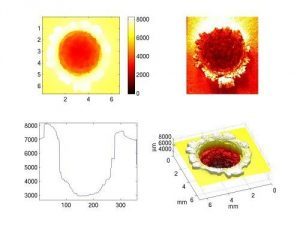
Surface tomography of a hypervelocity impact crater.
Profilometry of craters using coherence radar
- L. Kay, A. Gh. Podoleanu, M. Seeger, and C. J. Solomon, “A New Approach to the Measurement and Analysis of Impact Craters,” Int. J. Impact. Eng. 19, 739-753 (1997).
Imaging paintings
En-face OCT allows visualisation of under-drawings (Collaboration with the Nottingham Trent University, British Museum London and National Gallery, London). This was followed by a Leverhulme Grant.
- H. Liang, M. G. Cid, R. Cucu, G.M. Dobre, J. Pedro, D. Saunders, and A. Gh. Podoleanu, “Application of Optical Coherence Tomography to Examination of Easel Paintings,” Opt. Express 13, 6133 – 6144 (2005).