This project is suitable for a 1-year MSc in Physics. The project does not currently have funding attached, students must be able to fund the fees and their living costs either through their own funds or a scholarship. Current information on fees is available here.
Optical coherence tomography (OCT) is a non invasive, non touch imaging technology used to determine the structure of translucent objects with high resolution. It combines a high axial resolution with good tissue penetration. Whilst well established for retinal imaging, the equipment currently used in ophthalmology clinics does not provide reliable skin images, as needed in the investigation of periocular tumors and other skin diseases, where cellular resolution is needed. All progress in OCT has been directed towards improving the axial resolution, while the transversal resolution is still determined by the focusing power of the scanning lens. When trying to improve the lateral resolution, this comes against the OCT principle that collects all depths under a fixed focus.
Recently, the AOG has demonstrated a new OCT technology that can collect any number of en face images from as many depths required, in real time. The method is ideally suited to be used repetitively for different focus adjustments. Conventional OCT technology can also perform repetitive acquisitions under different focus positions, but the technology is time consuming, as conventional OCT delivers all volume, all depths for each acquisition, i.e. even the data from outside the focus, which is blurred. Our proprietary direct en face OCT method delivers en face images from the depths of interest only, which under tight focus may mean a single en face slice. No time is wasted on delivering information from outside the focus, as in conventional OCT, where after the acquisition, data from outside the focus is discarded. Each acquisition requires seconds, depending on the spectrometer speed. With our method, repetition of acquisition is sub-second, as no time is needed for selection and removal of data, only the necessary data is produced, that of the en face image for the focus position selected.
This immediately revolutionizes the OCT technology in terms of transversal resolution, as it is known that only the en face images from the focus present good transversal resolution. This opens the possibility to work under highly tight focus, as only confocal microscopy could do. Tight focus has not been used so far with conventional OCT for the reasons detailed above. Tight focus means that lateral resolutions as used in histopathology analysis becomes in this way available. We have proven in a recent publication [1] last year that when the number of repetitions exceeds 4, the time required by our direct en face OCT becomes less than the time required by the conventional OCT technology. This procedure is called Gabor. The project will combine for the first time the Gabor procedure with the Direct en face OCT method we have recently developed and patented[2], to acquire high resolution images of skin. Usually OCT images from the retina and commercial OCT instruments exhibit lateral resolutions of 15 microns x 15 microns. With Gabor and the Direct en face OCT we aim for lateral resolutions of at least 1 micron x 1 micron. The project also aims to improve the axial resolution beyond conventional values, to truly compete with histology. In depth the resolution is determined by the broadband optical source used. The larger the spectrum , the better the axial resolution. This is usually 5-10 microns.
To improve the axial resolution, we will take advantage of the recent work we performed with NKT Denmark, partner in a Marie Curie European Industrial Doctorate (EID) training site, 2014-2018. NKT is the world leader in supercontinuum, providing the broadest spectrum possible for OCT sources. Using supercontinuum to drive the OCT system, 1 micron becomes accessible in depth. With micron resolution both in the lateral and axial direction, exciting avenues are opened in transforming the OCT technology into a real time imaging procedure that can compete with histology in terms of resolutions.
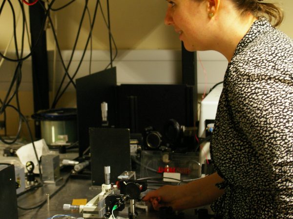
We aim to achieve some proof of concept, perfectly achievable. The student will perform optics assembly work and dedicated work on correlating data between high resolution OCT for histology and wider images collected in vivo. For the moment, all OCT groups recognise the limitations of the OCT in BCC diagnosis. We believe that by enhancing the resolution, OCT can compete with histology and provide better delineation of BCC, as well as open other diagnosis avenues, of skin diseases.
There is no deadline for the project – applicants will be assessed on a rolling basis – although please note any separate deadlines for scholarships or funding. For further information or informal enquiries, please contact Professor Adrian Podoleanu (A.G.H.Podoleanu@kent.ac.uk).