Imaging Centre for Life Sciences

Featured story

Imaging Centre for Life Sciences

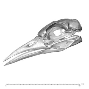
The Imaging Centre for Life Sciences conducts cutting edge scientific research across a broad spectrum of disciplines including palaeonotology, materials science, architecture, forensic science, biomedicine, space science, and archaeology. It also provides imaging services to business partners and other external users.

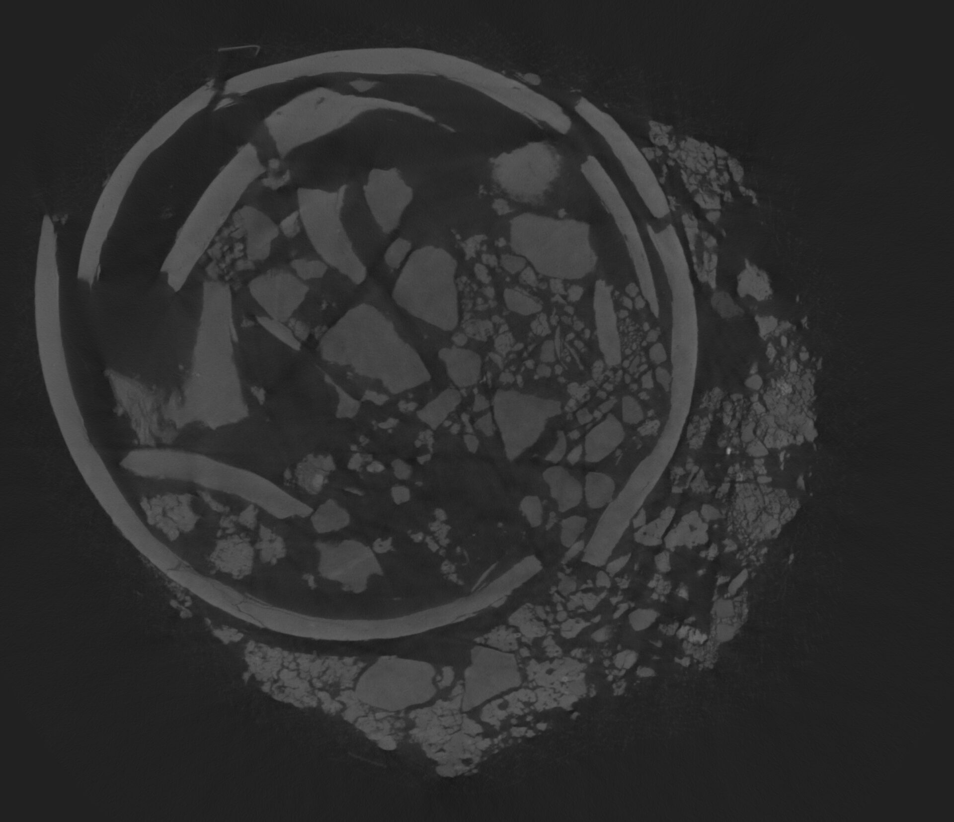
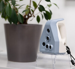
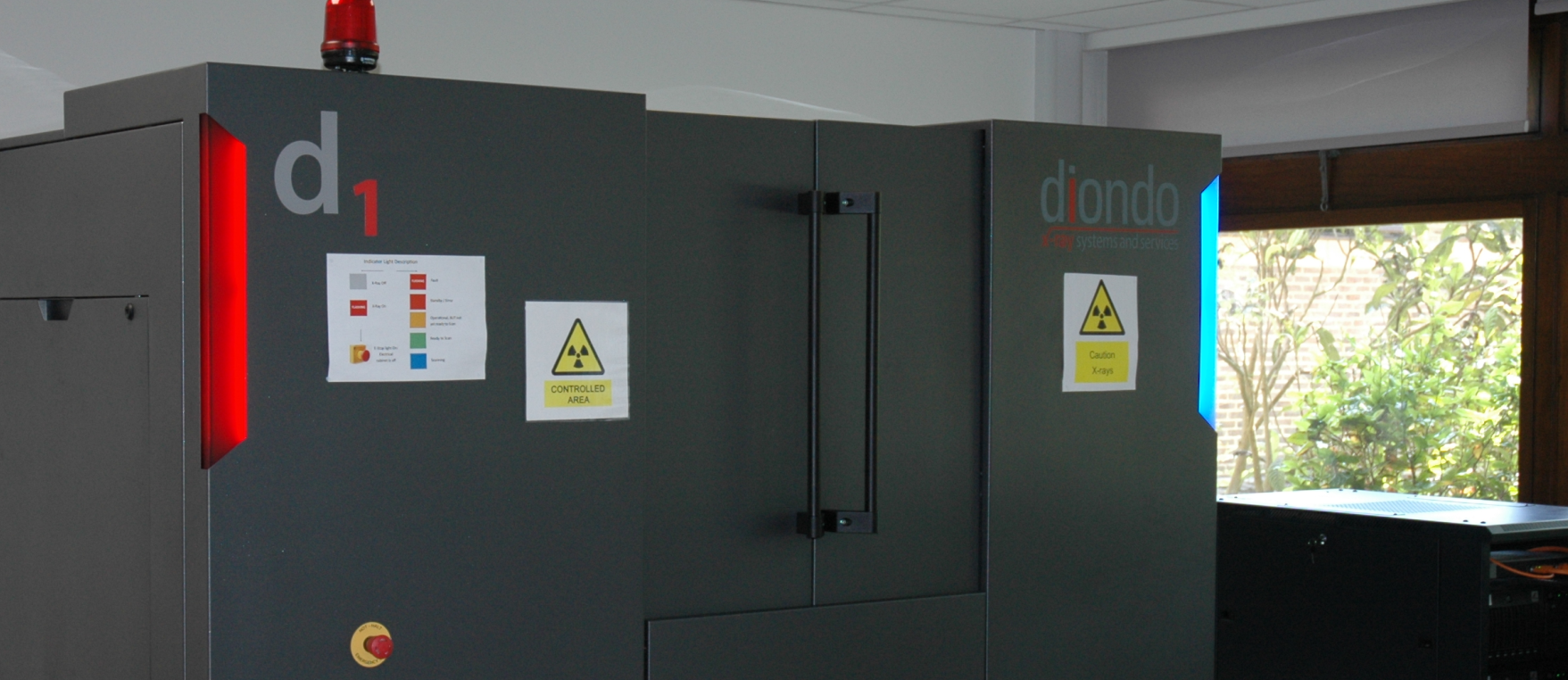
The Imaging Centre for Life Sciences (ICLS) houses a state-of-the-art Diondo d1 X-ray micro- computed tomographic (Micro-CT) scanner that can image and quantify the internal structure of most objects, down to a spatial resolution ranging from  ~5-150 micrometres (see below for precise specifications).

The centre images and measures objects for Commercial , Heritage , and Academic Research sectors.

Whether you are looking to understand the internal structure of your final product, measure improvement in your manufacturing process, ensure quality control, non-destructively see inside priceless archaeological artefacts, or need internal data for your scientific research, the ICLS can help.


Click on the links below to see how we can help you:

X-ray computed tomography is a non-destructive imaging technique that allows the visualization of the external and internal structure of objects. Through the creation of a series of radiographs a 3D volume of the scan is created which can then be visualized in order to examine parts of the object that differ in their density (or the degree to which they attenuate X-rays). Applications include but are not limited to anatomy and medicine, bone biology, forensic science, food technology, engineering, electronics, and geosciences.

Facilities

The Diondo d1 can scan objects with a spatial resolution up to ~5-150 micrometres depending on the size of the sample, with a scan volume up to 400 x 400mm and weighing up to 20kg.

Diondo d1 Micro-CT Scanner

Furthermore, our Artec Spider surface scanner is a photogrammetry based system that can be used to capture detail surface morphology of small and medium sized objects.

Artec Spider Scanner

Our technical staff can help you create a variety of visualization and digital objects suitable for analysis and 3D printing.

A surface model of a bird skull provided by Prof Jim Groombridge

Bird skull surface model

Services and Fees

We offer 3D visualization and analysis services to researchers and companies.

These can include:

  • CT-Scanning of objects
    • including sample mounting, technician time and reconstruction
  • Bespoke segmentation of images down to their constituent parts
    • including scientific researcher expertise
  • Visualisation of internal and/or microscopic structures
    • including 2D images, 3D volume images highlighting parts of interest
  • Analysis of  internal and/or microscopic structures
    • this can include quantitative measurements of the structures of interest


For pricing of your specific needs please do contact us:

Email

c.j.dunmore@kent.ac.uk

Call us

01227 824097