
We are investigating the local regulation mechanisms of important renal tubular transport mechanisms. These mechanisms are key to good health, and consequently, dysfunction of these regulatory mechanisms may account for a number of a number of poorly understood, and in some cases fatal, renal disorders.
We are particularly interested in the function of a variety (and abundance) of extracellular ATP-activated P2 receptors in the mammalian nephron. We have been investigating the molecular basis by which luminal P2 receptors alter epithelial Na+ channel (ENaC) activity in the distal nephron, using electrophysiological (patch clamp), molecular and imaging techniques on microdissected ‘split-open’ collecting duct tubules. We have identified a luminally-expressed P2 receptor that acts as a Na+ sensor and locally regulates ENaC activity and therefore may play a role in determining systemic blood pressure.
Investigation of local regulation mechanisms in renal tubular transport
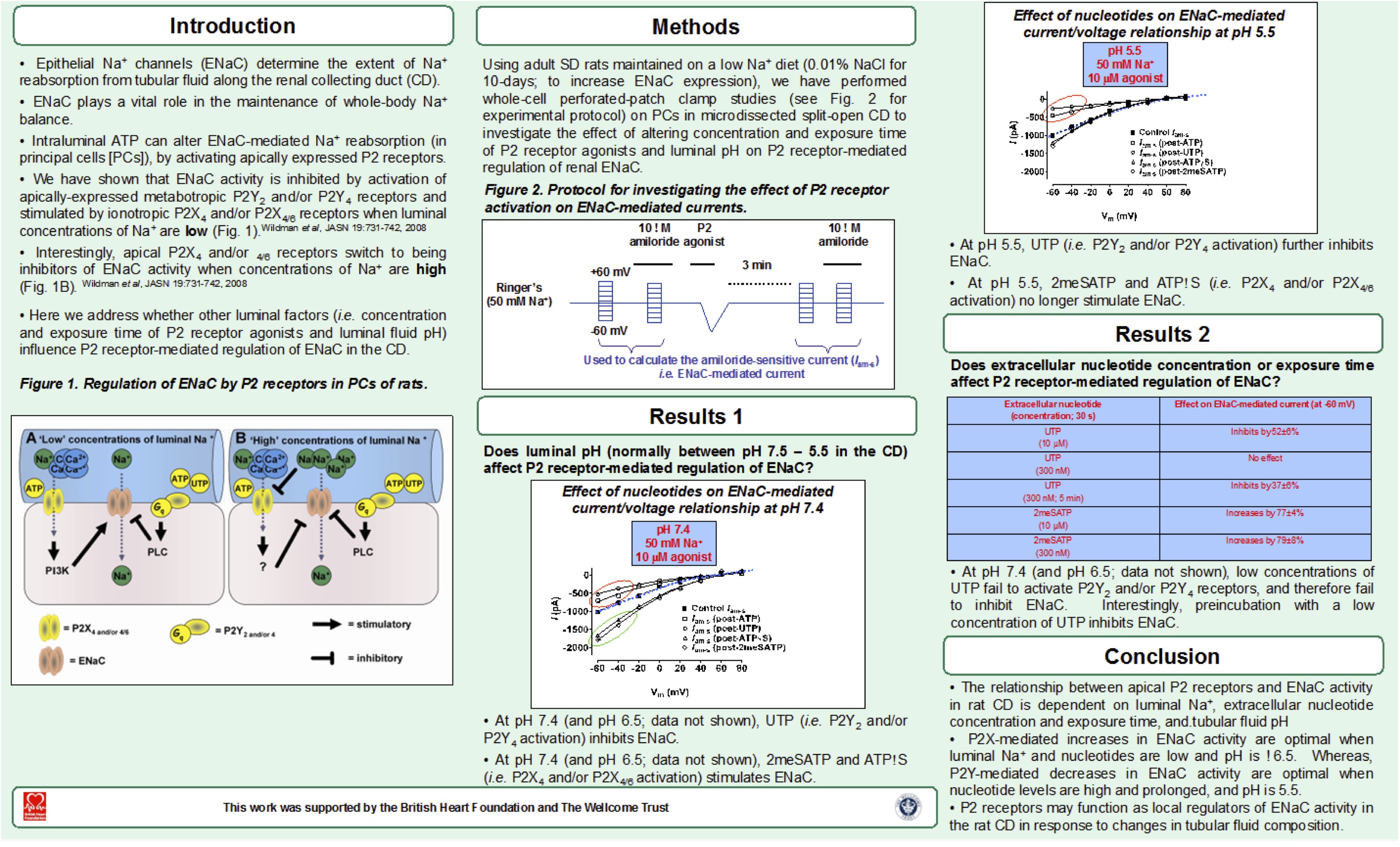
Recent poster presentation entitled ‘The complex nature of P2 receptor-mediated regulation of ENaC: dependence on Na concentration, nucleotide concentration and exposure time, and tubular pH’, by Scott S.P. Wildman and Sean G. Brown (Abertay)
We are interested in the regulation of renal medullary blood flow through the microvasculature.
We have developed a novel live kidney slice model to investigate the role of specialised smooth-muscle-like cells, termed pericytes, in the regulation of in situ vasa recta capillary diameter. Since the renal medulla is relatively inaccessible in vivo the slice model provides an ideal environment in which to characterise endogenous modulators of vasa recta pericyte contractility. Using the slice model we are also investigating tubulovascular cross-talk and mechanisms of drug-induced nephrotoxicity.
Investigation of the regulation of renal medullary blood flow
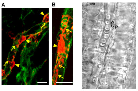
Identification of vasa recta pericytes.
Pericytes (red) are composed of cell bodies (arrowheads) and ‘claw-like’ processes (arrows) that extend from the cell body to run along and wrap around the capillary (Crawford et al., 2012). Pericytes are located at regular intervals along vasa recta capillaries (green). Kidney slices from NG2 Ds-Red Bac transgenic mice in which pericytes express fluorescent Ds-Red (red; b) have been used in experiments to aid the identification of vasa recta pericytes (Lydon et al., 2012).
We are interested in urothelial cell signalling (in particular extracellular ATP signalling) in health and disease. Abnormal urothelial cell signalling has been linked to a number of bladder disorders, including Overactive Bladder syndrome (OAB).
OAB is estimated to affect 100 million adults worldwide. Symptoms include a frequent, sudden and urgent desire to pass urine, incontinence and in some cases chronic pain. Current treatments are not ideal, being hindered by side effects. We are investigating a low-level inflammatory response that affects the urothelium in a significant subset of OAB patients; this inflammatory response may account for their symptoms.
Investigation of urothelial cell signalling in health and disease
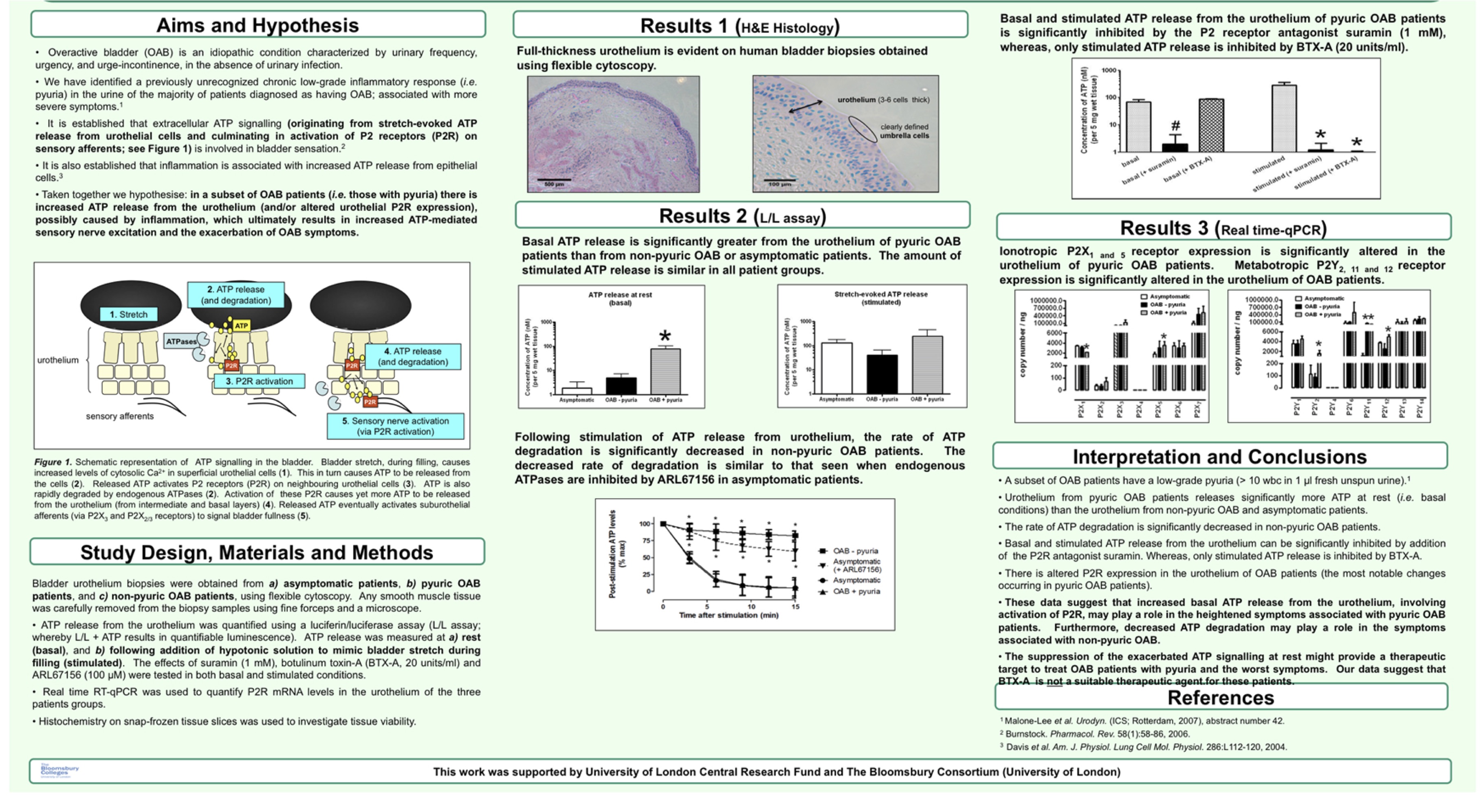
Recent poster presentation entitled ‘Altered ATP signalling in urothelium of OAB patients with exacerbated symptoms ’, by Alberto Contreras-Sanz, Scott S. P. Wildman et al.