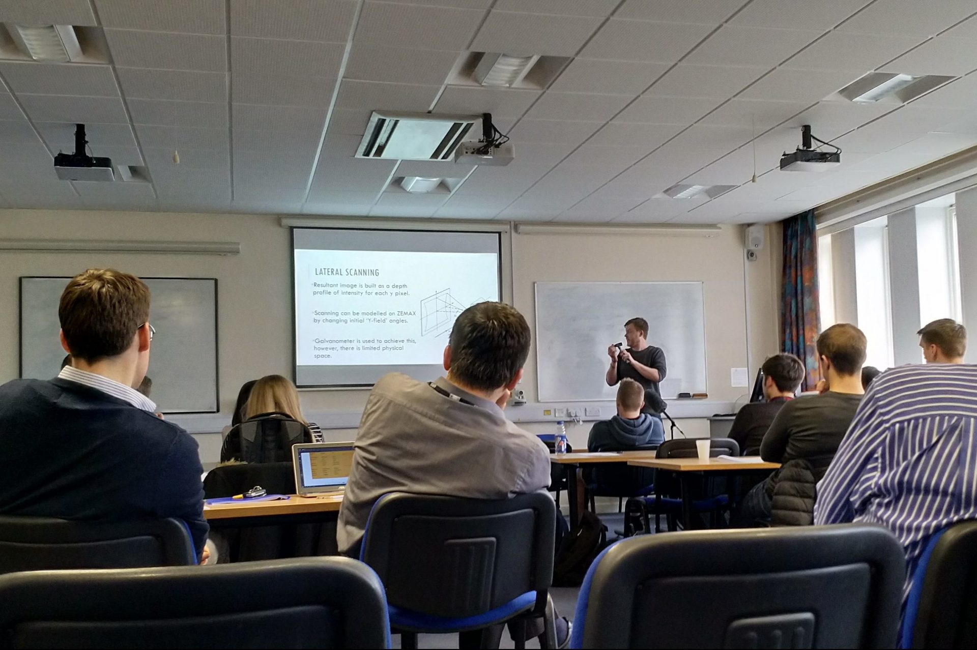
On the afternoon of Tuesday 27th March, four SPS MPhys students who carried out their fourth year projects with the AOG have given their final presentations to an audience consisting mainly of their peers and academic/research staff from the school. The projects were quite diverse, ranging from white light interferometry to endomicroscopy, with optical design using ZEMAX and novel multi-modal imaging methods also making an appearance.
The full title and author of each presentation were as follows:
-
Dispersion in Optical Coherence Tomography Systems, Fiona Bairstow (supervised by Prof Adrian Podoleanu)Complex Master Slave Interferometry (CMSI) is a novel imaging method developed to decode an output spectrum from an interferometer without dispersion negatively affecting the final image. CMSI adequately compensates for dispersion in the optical system and nonlinearities in the spectrometer using two mathematical functions, called g and h respectively. The way in which dispersion, due to SF6 glass and water objects, affects these functions has now been investigated. The function g was found to be unchanged as expected. The function h did change due to both objects, however, not by a large enough amount to allow for the calculation of the correct value of group velocity dispersion. Therefore, it was concluded that CMSI is currently not capable of compensating for all dispersion introduced by the object to be imaged, and additional mathematics may be needed in order to achieve full compensation.
-
The characterisation and optimisation of a multimodality optical coherence tomography and photoacoustic tomography imaging instrument, Haidar Awad (supervised by Dr Adrian Bradu)A first aim of the project was to investigate and compare the characteristics of an optical coherence tomography (OCT) imaging instrument equipped with either the SuperK Compact (SKC) or the OptoSpeed SA SLED (SLED) laser sources operating in the near infrared (NIR). The OCT setup and a photoacoustic tomography (PAT) channel were part of a multimodality imaging instrument, where the OCT was operating in NIR while the PAT in visible (VIS). A second goal of the project was to devise the OCT channel to operate in the same spectral range (VIS) as the PAT channel. Characteristics were obtained by conducting sensitivity tests and comparing the quality of B-Scan images, axial resolution, power, and noise levels produced by each of the two optical sources. To allow for the OCT and PAT to operate in the visible spectral range a new spectrometer, to detect the optical signal and a new reference arm of the interferometer had to be built. Results showed that both optical sources had a very similar 3dB bandwidth imaging axial depth despite having a greater signal amplitude for the SKC, suggesting more noise in backscattered photons for the SKC. The SKC produced a baseline noise level that was 14.38 times greater than the SLED. Although, the SKC had a better axial resolution. The SKC B-Scans showed this huge difference in noise however, the features within the sample were sharper due to the better axial resolution. A total sensitivity of 76.42 and 50.33 was measured for the NIR SLED and SKC respectively. With regards to the PAT channel, an increase in photoacoustic signal sensitivity was observed as the spectral bandwidth of the SKC was increased.
-
The Design of an Otoscope Attachment for the in-vivo Imaging of the Middle Ear, Liam Sexton (supervised by Dr George Dobre)
In this talk, I will discuss the process of designing an attachment that will fit on a commercial otoscope. A 1 to 1 correspondence has been shown between the bacterial biofilm growth and the development of an ear disease known as Otitis Media, which affects 1 in 3 children worldwide. Currently, the gold standard for the detection of Otitis Media is myringotomy – a highly invasive procedure which involves the surgical incision of the ear drum. For this reason, the attachment uses Optical Coherence Tomography for non-invasive depth imaging of the middle ear and thus the detection of biofilms. The design produced has been done using a group of ray-tracing optical design applications known as ZEMAX and has been shown to achieve a lateral resolution of 7.85 μm for a 380 μm field of view in the image plane. Further analysis has shown that increasing the field of view to 920 μm produces an off-axis resolution of 18.12 μm, allowing a clinician to achieve a greater field of view at the expense of image resolution.
-
Modelling Structured Illumination Endomicroscopy, Connor Stroud (supervised by Dr Michael Hughes).Endomicroscopy is a process of taking histology style images inside the human body in real- time, typically by using an optical fibre bundle. Structured illumination is a process wherein a pattern of lines, alternating between black and white, is used to illuminate a specimen. The lines are shifted by a third of their phase to the right and the specimen is illuminated again. Images are taken after each illumination. Three images are taken in total and are used to gain an optically sectioned image. In this talk I will be describing my project, which is based on developing a model in which a specimen is observed using an optical fibre bundle. The projects main aims were to apply structured illumination to the model, to find the width of the lines within the pattern which creates an image with the optimal axial resolution with a good noise to signal ratio, and to recreate this model experimentally to validate the results found.
All talks were well-received and attracted pertinent questions. We congratulate all four students for their efforts and wish them all the best in their future endeavours!